Fluoroscopy

What is Fluoroscopy: An Informative Guide
Fluoroscopy is a medical diagnostic technique that uses X-rays to generate real-time images of the inside of the body. It is commonly used by radiologists and other medical professionals to diagnose a range of conditions. It is a safe and non-invasive procedure that is often performed in an outpatient setting. If you have been referred for a fluoroscopy examination, you may be wondering what it is, how it works, and what to expect. So, in this blog, we will discuss everything you need to know about fluoroscopy, types of fluoroscopy, when do you need it, how to prepare, and what to expect before, during, and after the exam.
Types of Fluoroscopy
There are different types of fluoroscopy that are used to diagnose various conditions. Some of the most common types of fluoroscopy include:
1. Barium Swallow: It uses a thick liquid called barium to coat the lining of the esophagus.
2. Upper GI Series: It is used to evaluate the upper portion of the digestive system, including the esophagus, stomach, and small intestine.
3. Lower GI Series: It is used to evaluate the lower portion of the digestive system, including the colon and rectum.
4. Arthrography: It is used to diagnose joint problems by injecting a contrast dye into the joint.
When Do You Need It
Your doctor may recommend a fluoroscopic examination if you are experiencing symptoms such as abdominal pain, difficulty swallowing, joint pain, or bowel problems. It can be used to diagnose problems such as ulcers, tumors, hernias, or blockages. It is important to follow your doctor’s instructions on when and why you need a fluoroscopic examination.
How to Prepare
The preparation for a fluoroscopic examination varies depending on what part of the body is being examined. For example, if you are having a barium swallow or upper GI series, you may need to fast for several hours before the exam. You may also be asked to avoid certain foods or medications. Your doctor will provide you with specific instructions on how to prepare for your exam.
What to Expect Before, During, and After the Exam
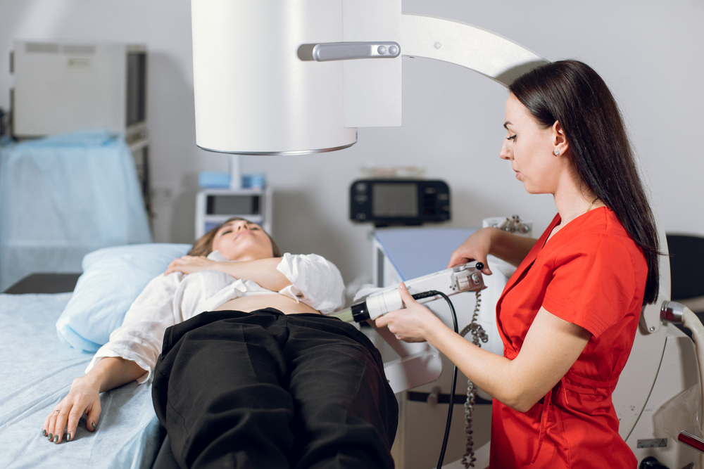
Before the exam, you will be asked to change into a hospital gown and remove any metal objects or jewelry. You may also need to remove dentures, depending on the type of exam. During the procedure, you will lie on a table while the X-ray machine generates real-time images of the inside of your body. The procedure usually takes 30 to 60 minutes, depending on the type of exam. After the exam, you will be able to go home. You may experience mild discomfort or diarrhea, depending on the type of exam.
Fluoroscopy is an important diagnostic tool that can help diagnose various medical conditions. It is a safe and non-invasive procedure that is often performed in an outpatient setting. If you have been referred for a fluoroscopy examination, it is important to follow your doctor’s instructions on how to prepare and what to expect before, during, and after the exam. While the exam may be uncomfortable or inconvenient, it can provide valuable information that can help diagnose and treat your condition. So, if your doctor has referred you for fluoride testing, don’t be anxious, it is a simple and a routine test.

Less Movements Just as Effective In Ultrasound for Pancreatic Biopsies
A new Japanese study shows that fewer to-and-fro movements during an endoscopic ultrasound-guided fine-needle biopsy (EUS-FNB) are equally effective when performing a pancreatic tumor biopsy. Gastrointestinal Endoscopy published

Increased Use of CCTA Cost Effective for Diagnosing Coronary Artery Disease
Radiologic screening opportunities are on the increase in the field of cardiology. A new retrospective study examined the results in the United Kingdom of

New FDA Recommendation for Women with Dense Breast Tissue
The U.S. Food and Drug Administration updated their mammography standards in early March to require imaging providers to inform women that they have dense
