If you suffer from joint pain or mobility issues, your doctor may recommend an arthrography test. Arthrography is a diagnostic medical imaging technique that uses contrast dyes and X-rays or MRI to visualize the inside of a joint. This test can help identify joint injuries, diseases, or abnormalities in the bone and soft tissue. In this post, we’ll explain the different types of arthrography, discuss when it’s necessary, how to prepare for it, and what to expect during and after the exam.
1. What is Arthrography?
Arthrography is a medical imaging procedure that involves the injection of contrast dye into a joint. The contrast dye helps highlight the joint structure and surrounding tissue, allowing doctors to obtain accurate images of the joint’s interior. Arthrography can be done with X-rays (known as Fluoroscopic Arthrography) or MRI (known as MR Arthrography) to produce detailed images that aid in the diagnosis of joint injuries, arthritis, and other abnormalities.
2. What are the Different Types of Arthrography?
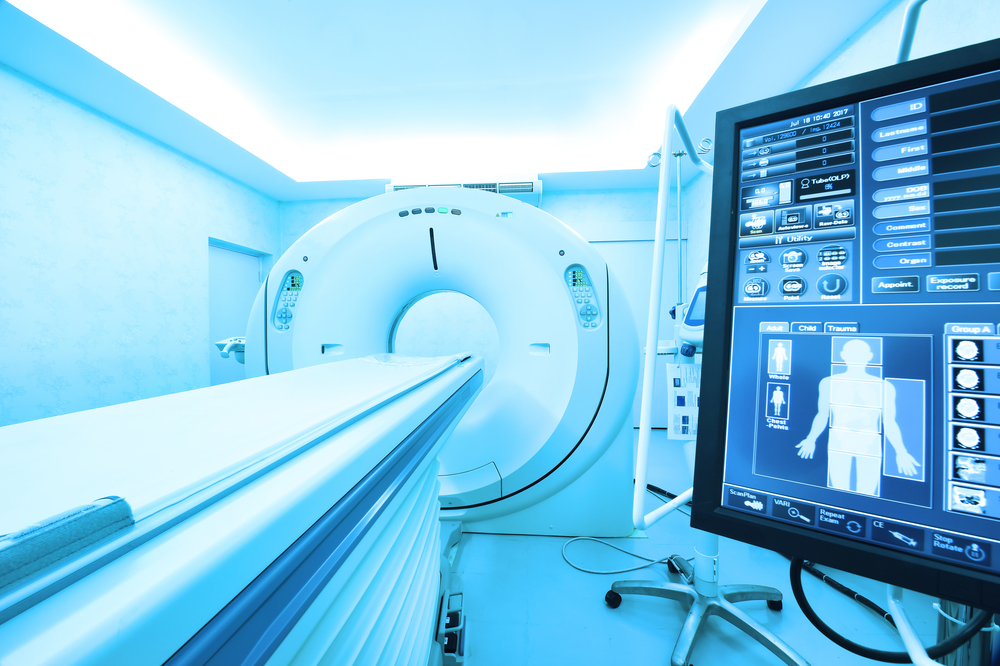
Two types of Arthrography are typically performed: Fluoroscopic Arthrography and MR Arthrography. Fluoroscopic Arthrography involves the use of a fluoroscope, a machine that emits a constant X-ray beam and allows a radiologist to view the joint in real-time. With MR Arthrography, an MRI scanner is used rather than X-rays, which provides a more detailed picture of the joint. Both types of arthrography have their distinct advantages depending on the joint in question and the health status of the patient.
3. When Do You Need Arthrography?
Arthrography may be recommended if you’re experiencing any unexplained joint pain or discomfort, or if doctors suspect any joint injuries, inflammation, or other abnormalities. You may be a candidate for arthrography if you have:
– Limited range of motion
– Pain in the joint that does not subside
– Swelling or stiffness
– Chronic joint inflammation
– Loose or detached bone or cartilage fragments
4. How to Prepare for Arthrography?
Before arthrography, your radiologist will provide you with instructions on how to prepare, depending on the type of test you’re undergoing. Generally, instructions may include:
– Fasting for a certain amount of time before the test
– Informing the healthcare professional administering the exam of any allergies or medical problems
– Wearing loose, comfortable clothing and avoiding jewelry
– Arranging for someone to take you home post-exam, in case any sedation is necessary
5. What to Expect Before, During, and After Arthrography?
One acknowledged advantage of arthrography is that it typically does not require sedation. During Fluoroscopic Arthrography, the physician will often start by injecting a local anesthetic into the joint of interest, which may cause a slight sting, similar to a bee sting, for a few seconds. Then an injection of contrast medium is given into the joint. The radiologist will then take a series of X-ray images to produce detailed images of the joint or use an MRI machine. The entire test usually takes anywhere from 30 to 60 minutes. After the test is complete, you can typically resume normal activities immediately.